Professional USB microscope · Dermatology
Clinical Dermatology
Magnified surface viewing and image documentation for qualified dermatology workflows.
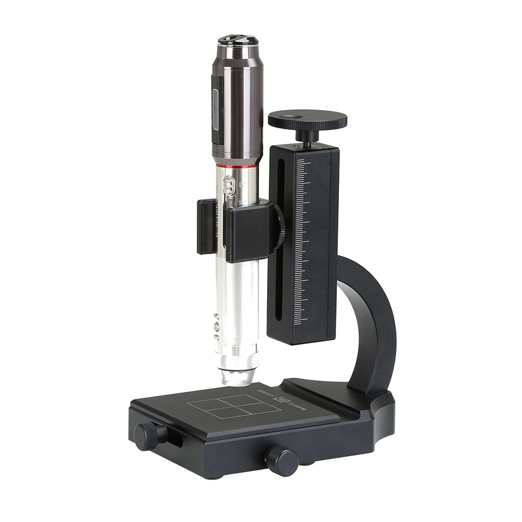
- Four quick magnification levels40x, 100x, 180x and 250x with the general inspection end-cap
- 3Pi narrow-tip end-cap100x access to small or recessed areas that remain in direct line of sight
- Quality optics, autofocus + Full HDCorning® Varioptic® liquid lens, photos and 30fps video
- Professional European supportShips from Spain with a two-year warranty
Overview
Dermatology workflows benefit from consistent visual documentation of the skin surface. Smart G-Scope provides a portable magnified-view and image-capture tool for qualified professionals.
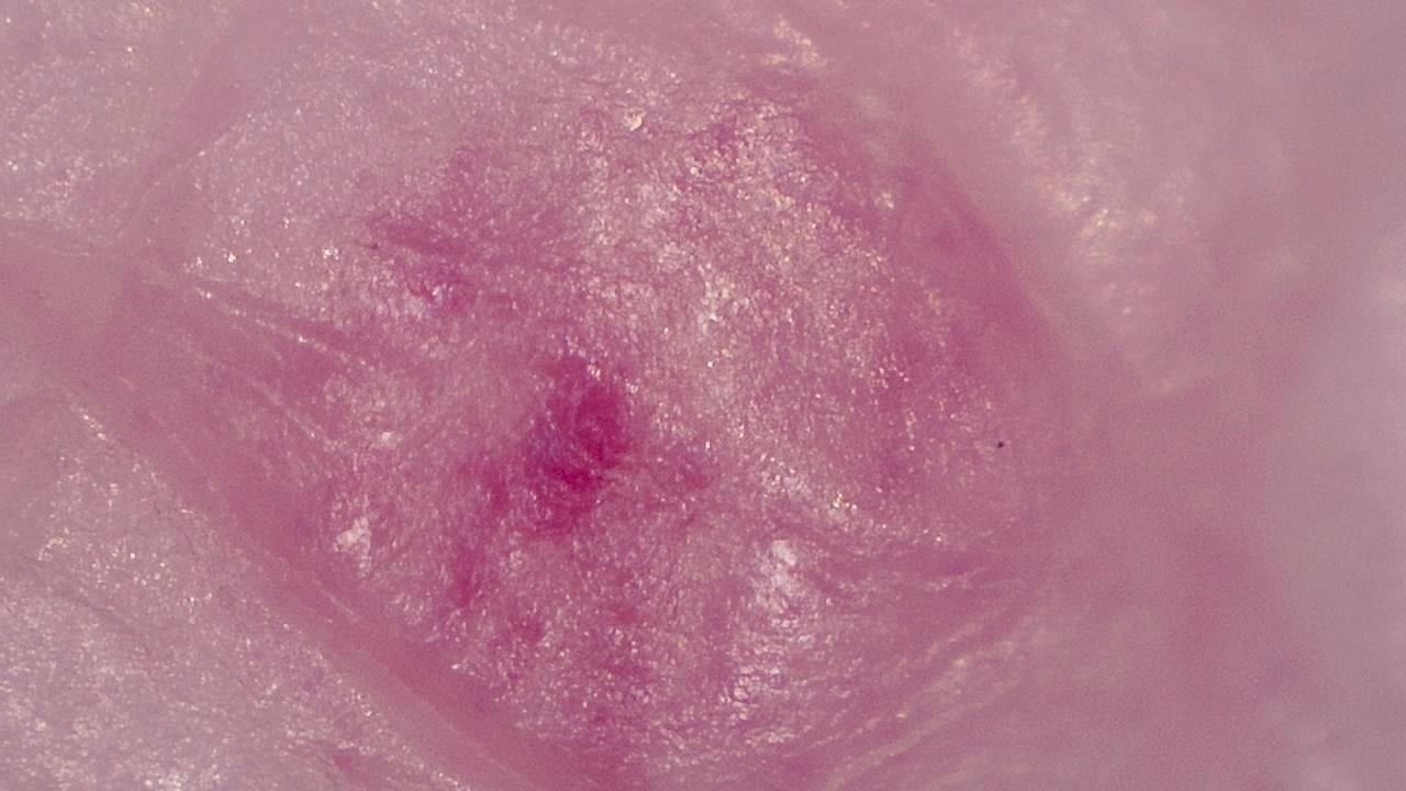
Its variable magnification can record visible surface pigmentation, vascular appearance, borders, and texture. It does not image beneath the surface or diagnose a lesion, and the clinical meaning of an image must be assessed by a qualified professional.
The integrated software enables photo and video capture to support longitudinal lesion follow-up, communication with other specialists, and clinical documentation aligned with healthcare quality standards.
Key benefits
- Visualization of dermal structures with up to 250× magnification
- High-resolution photographic documentation for the clinical record
- Longitudinal lesion follow-up through visual comparisons
- Portability for in-clinic and home visits
Image gallery
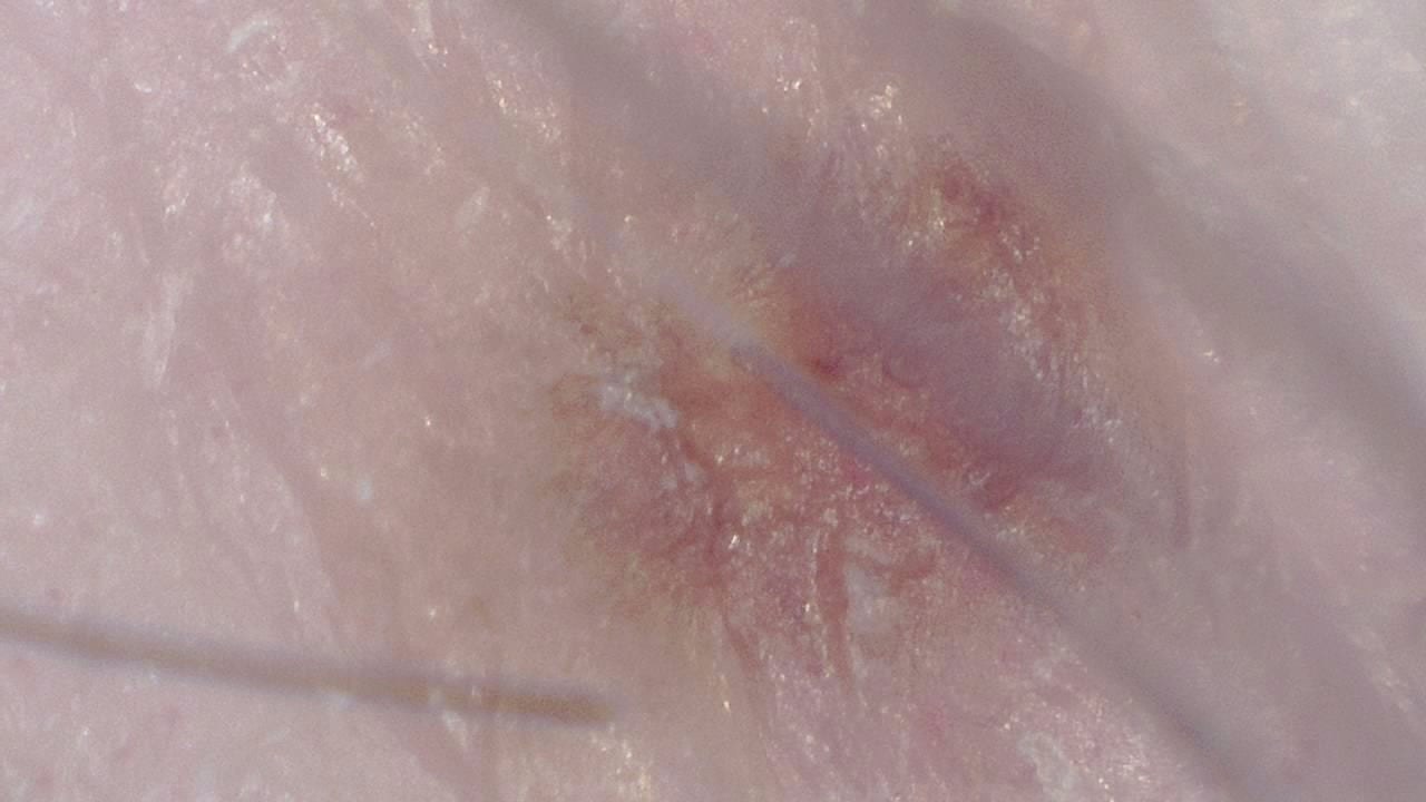
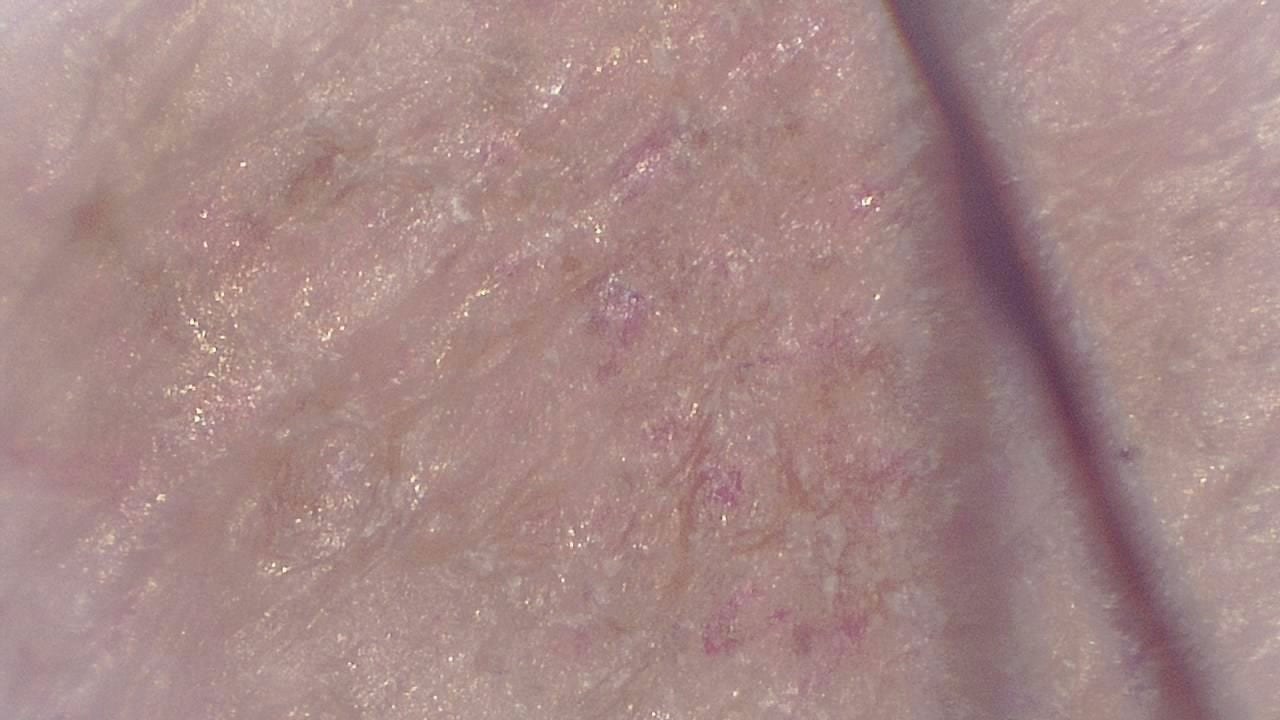
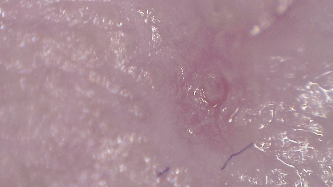
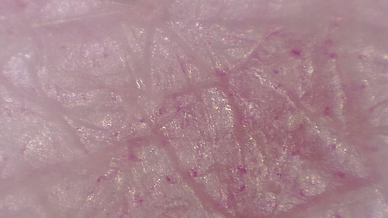
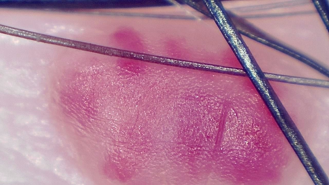
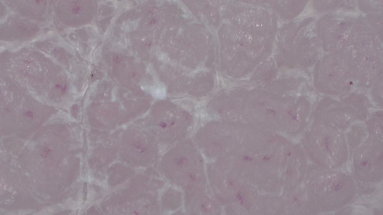
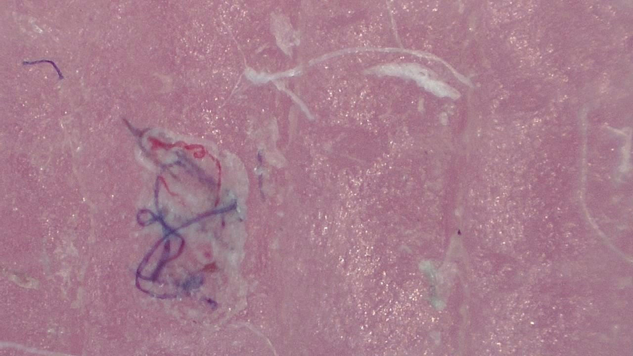
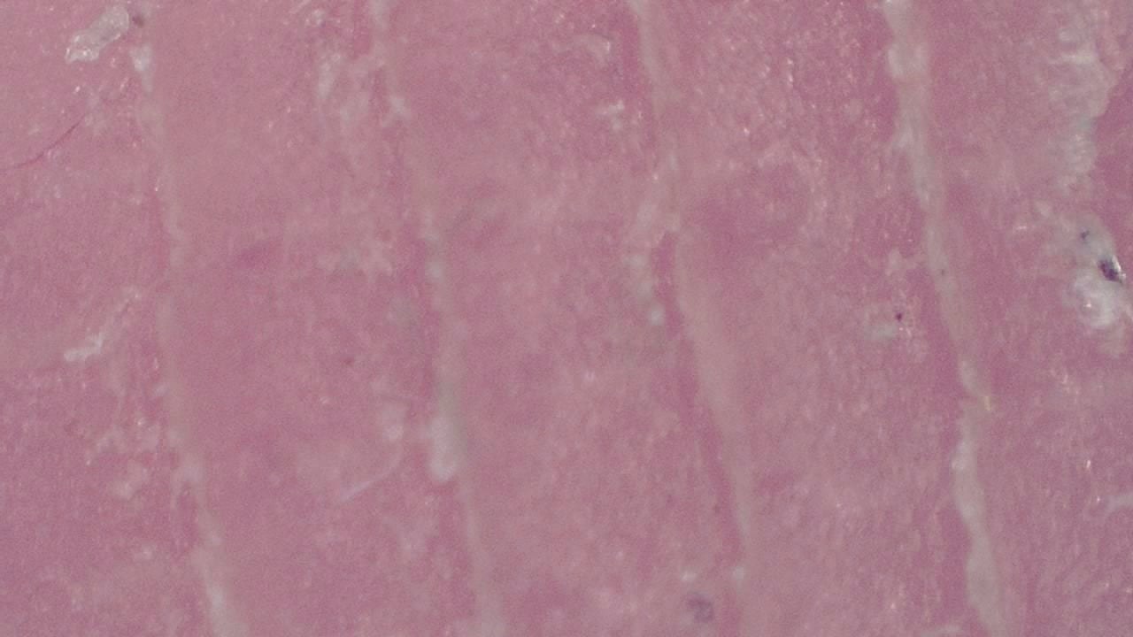
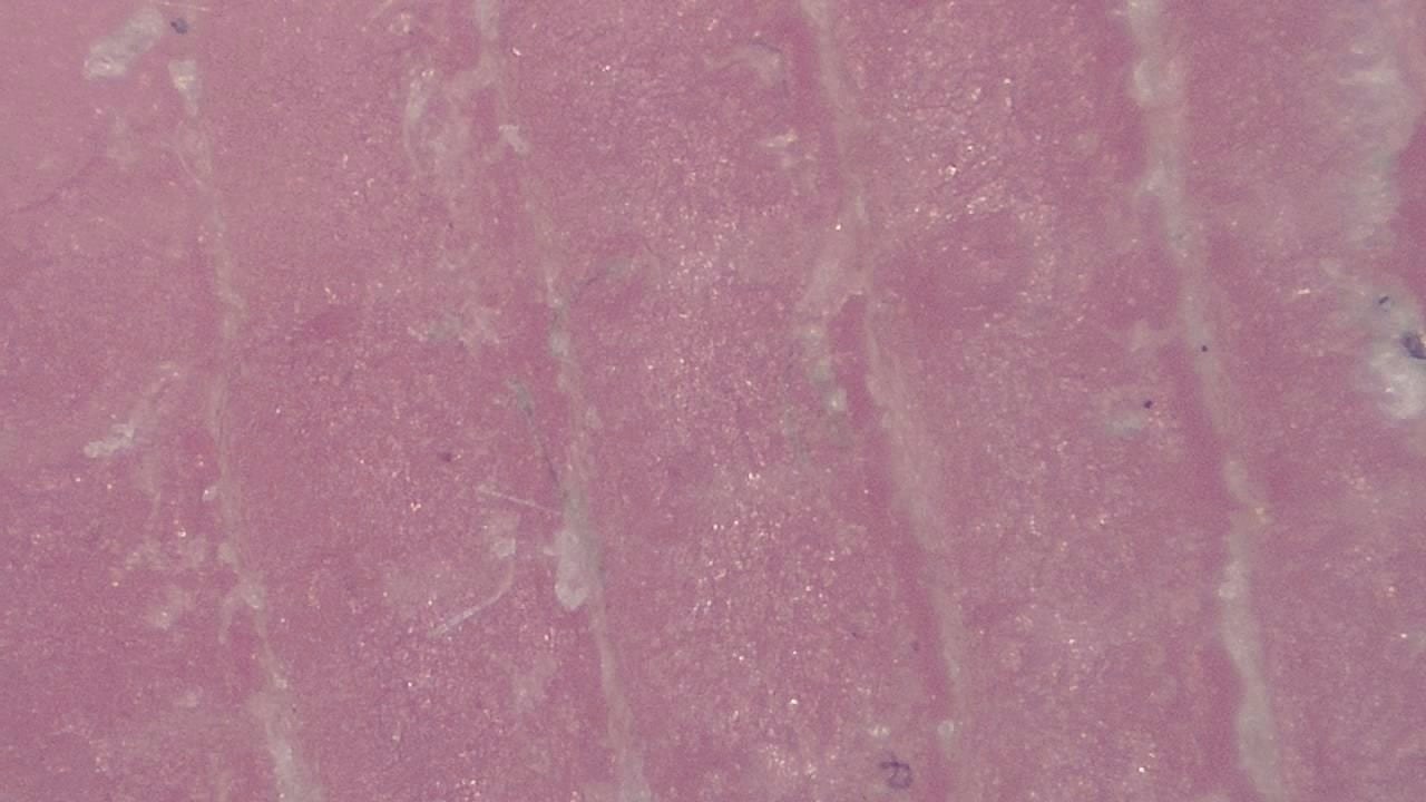
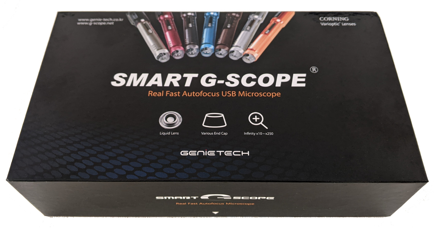
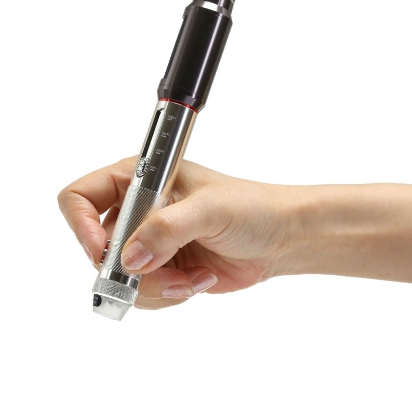
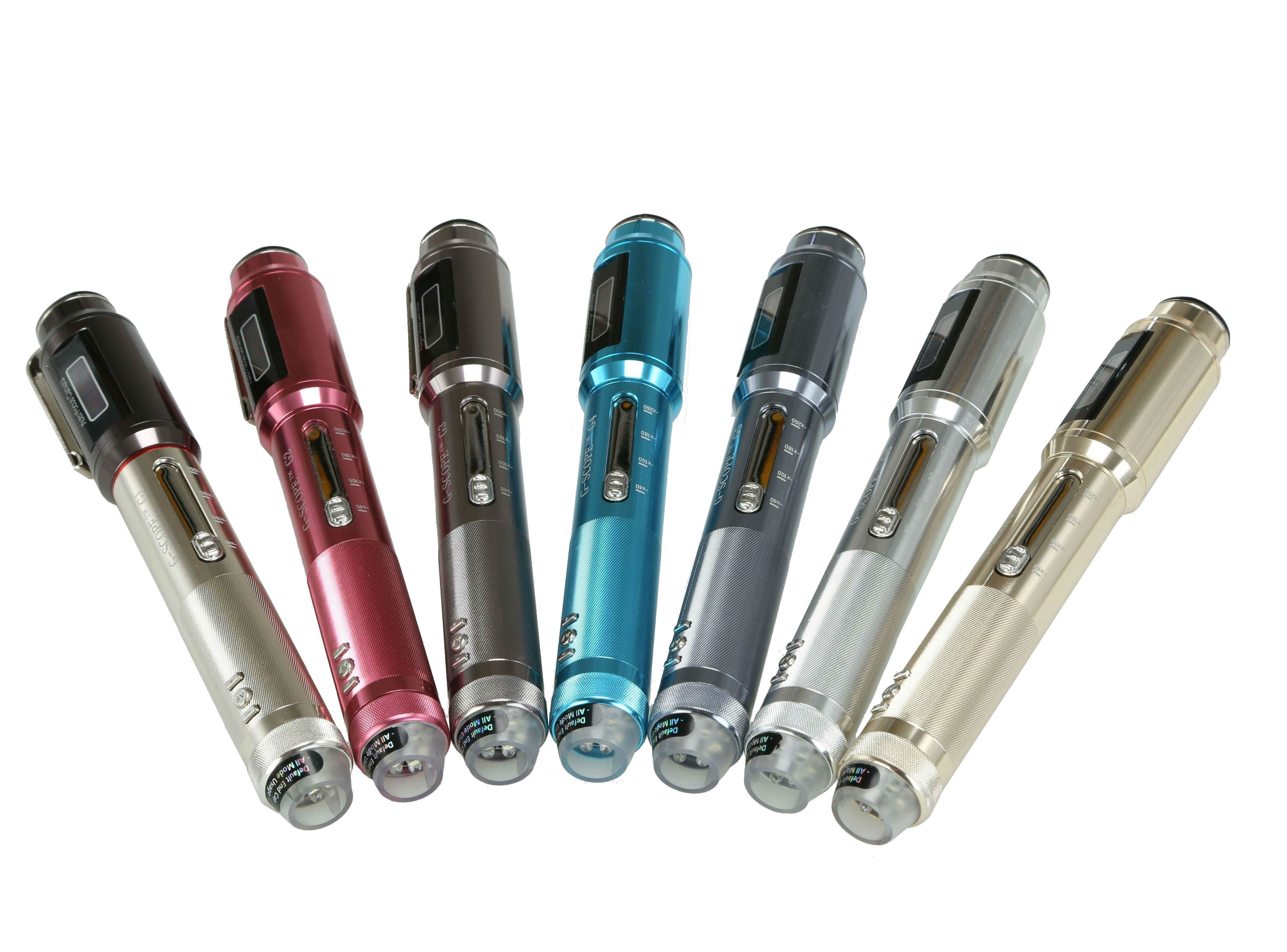
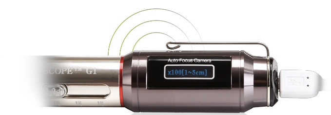
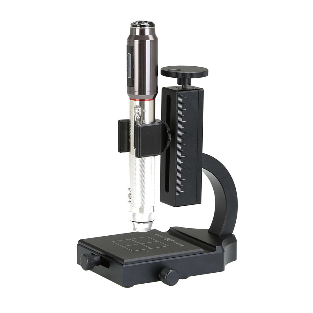
Click any image to enlarge
Practical examples
Atypical nevus follow-up
A dermatologist uses Smart G-Scope to document a lesion under consistent conditions during follow-up. Comparative images provide an additional visual record, while clinical assessment determines whether dermoscopy, biopsy, or referral is needed.
Contact dermatitis assessment
In an allergy clinic, the specialist documents visible texture and vascular appearance under consistent conditions. Treatment decisions use the complete clinical assessment rather than the microscope images alone.
Limitations & best practices
- Images are indicative and do not replace histopathological diagnosis.
- Keep the lens clean to avoid artifacts in captures.
- Adjust illumination according to the patient’s skin phototype.
- Comply with GDPR when storing patient images.