Professional USB microscope · Microbiology
Basic Microbiology
Observation of colonies, microbial structures, and preparations for teaching and basic research.
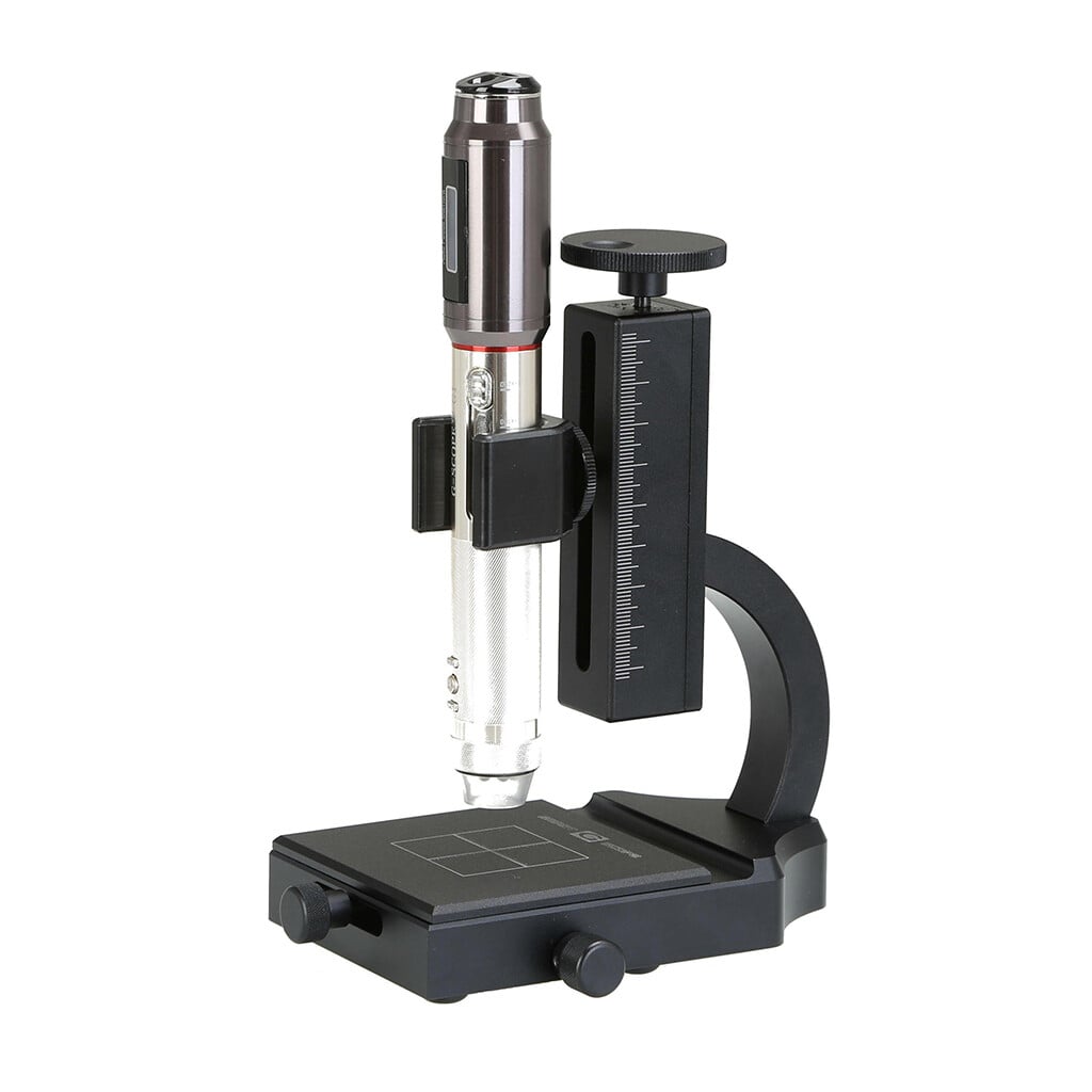
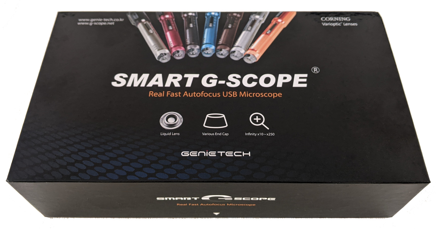
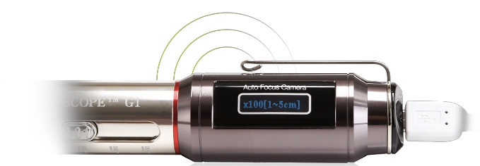
- Four quick magnification levels40x, 100x, 180x and 250x with the general inspection end-cap
- 3Pi narrow-tip end-cap100x access to small or recessed areas that remain in direct line of sight
- Quality optics, autofocus + Full HDCorning® Varioptic® liquid lens, photos and 30fps video
- Professional European supportShips from Spain with a two-year warranty
Overview
Although Smart G-Scope does not reach the magnification of a laboratory optical microscope, it is useful for basic and macroscopic microbiology observations. It can help examine bacterial and fungal colonies on plates, yeast preparations, filamentous algae, and fungal structures.
In microbiology teaching, the device helps students become familiar with colony morphology and capture images for lab reports, complementing the traditional microscope.
In low-budget research or citizen science projects, Smart G-Scope is an accessible option for documenting environmental samples and sharing findings online.
Key benefits
- Observation of colonies and macroscopic morphology
- Culture documentation for reports
- Image capture of wet mounts
- Accessible tool for microbiology education
- Citizen science and outreach projects
Image gallery
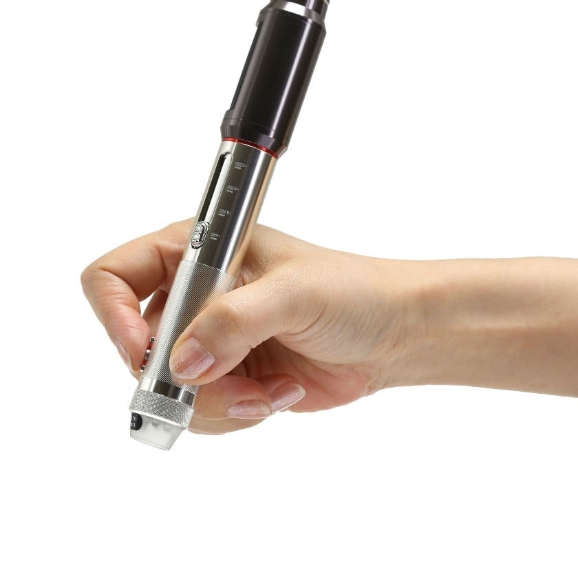
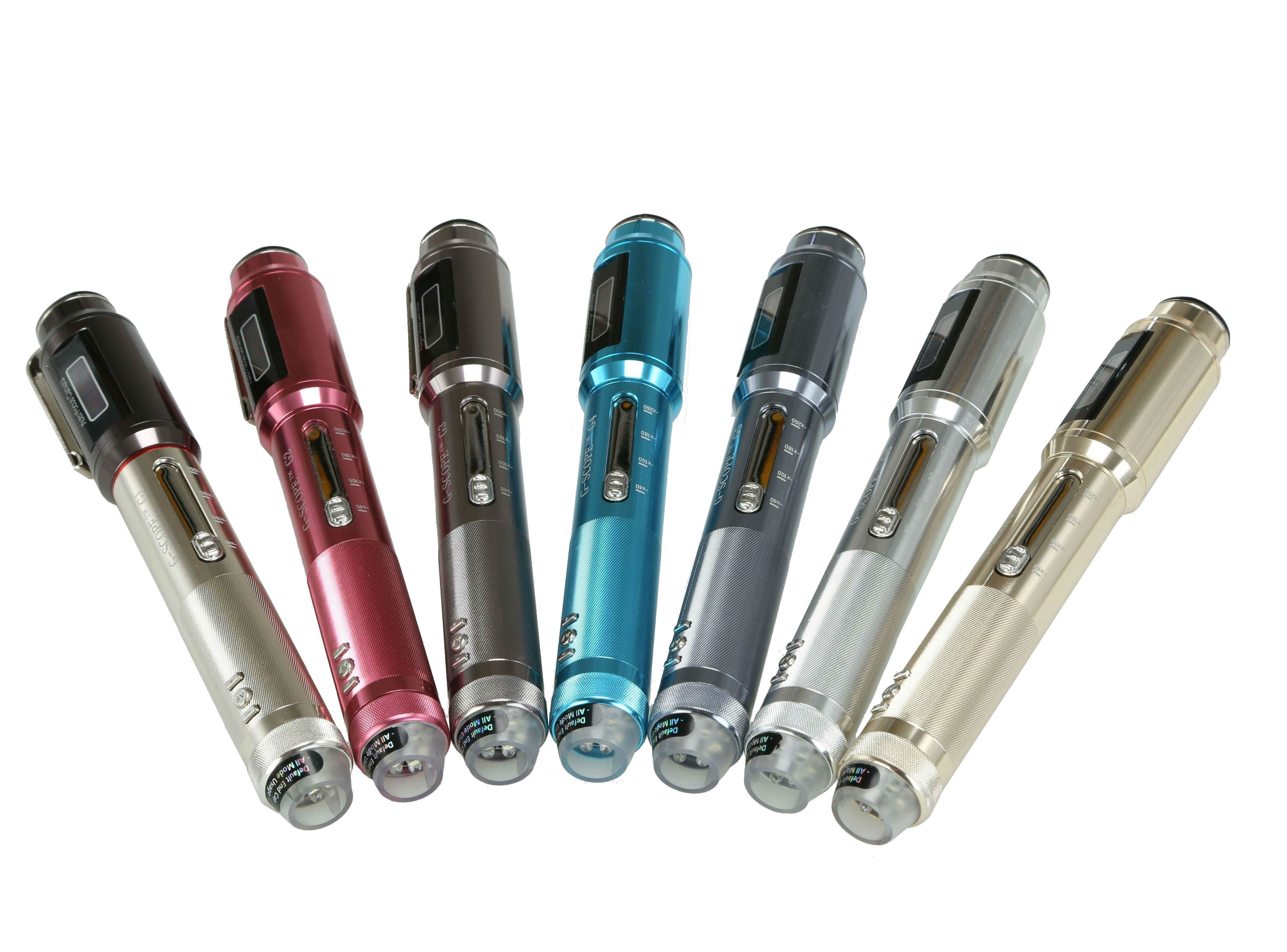
Click any image to enlarge
Practical examples
Teaching lab practice
Biotechnology students document colonies from different bacterial strains grown on selective media, comparing morphology and pigmentation using Smart G-Scope.
Citizen science project
A nature group documents visible algae and larger structures in prepared pond-water samples. Identification of microorganisms requires suitable microscopy and expert methods.
Limitations & best practices
- Insufficient magnification for detailed observation of individual bacteria.
- Staining and immersion microscopy require laboratory equipment.
- Follow biosafety protocols when handling potentially pathogenic samples.