Professional USB microscope · Veterinary
Veterinary Dermatology
Magnified viewing and documentation of accessible skin and coat surfaces in veterinary workflows.
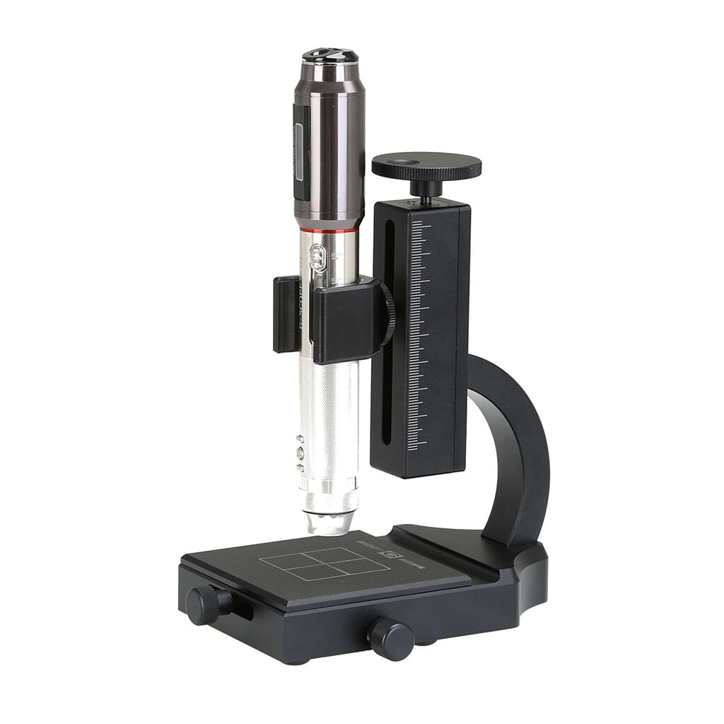
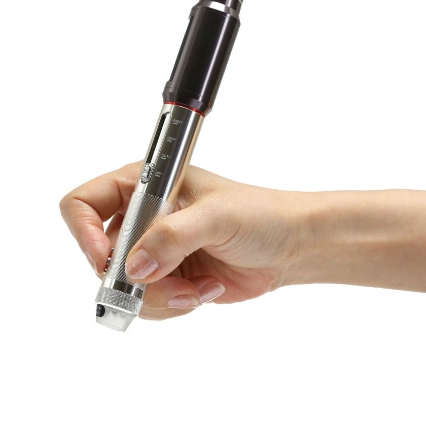
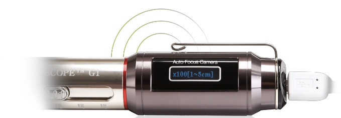
- Four quick magnification levels40x, 100x, 180x and 250x with the general inspection end-cap
- 3Pi narrow-tip end-cap100x access to small or recessed areas that remain in direct line of sight
- Quality optics, autofocus + Full HDCorning® Varioptic® liquid lens, photos and 30fps video
- Professional European supportShips from Spain with a two-year warranty
Overview
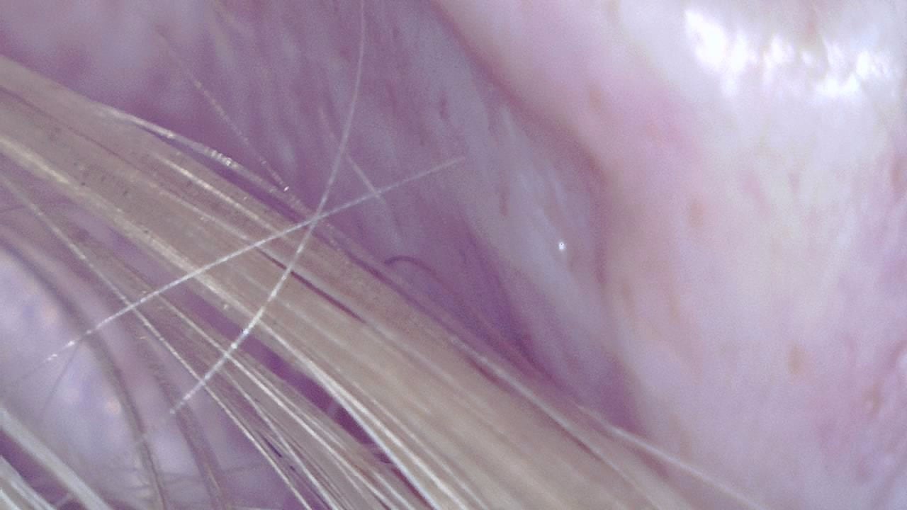
Veterinary dermatology brings unique challenges due to the diversity of species, coats, and skin conditions. Smart G-Scope enables veterinarians to examine the skin and hair of dogs, cats, horses, birds, and exotic animals with enough detail to support clinical assessment.
It can document visible coat, hair-shaft, skin-surface, and larger ectoparasite features when these are optically accessible. Fungal, allergic, follicular, and parasitic conditions still require veterinary examination and appropriate sampling or laboratory tests.
Visual documentation supports treatment follow-up and improves communication with owners, who appreciate being able to see their pet’s progress.
Key benefits
- Magnified documentation of visible skin, coat, and larger ectoparasite features
- Assessment of coat quality and follicular density
- Longitudinal documentation of dermatitis and alopecias
- Effective visual communication with owners
- Use in clinic, home visits, or farms
Image gallery

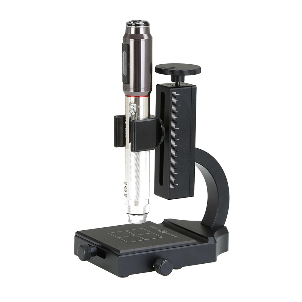
Click any image to enlarge
Practical examples
Veterinary skin-surface documentation
A veterinarian captures magnified images of accessible skin and coat features in a dog with pruritus. Diagnosis and treatment are based on the complete examination and appropriate confirmatory tests.
Feline atopic dermatitis follow-up
In a cat with chronic allergic dermatitis, the microscope helps document skin improvement after dietary adjustments, showing the owner reduced erythema and excoriations.
Limitations & best practices
- Animal immobilization may require assistance or mild sedation.
- Interpret findings in the context of species and breed.
- Confirm parasitological diagnosis with skin scrapings when needed.